Bio6
TRANSPORT SYSTEM IN MAN-CIRCULATORY SYSTEM
Circulatory system(man)=system of tubes(blood vessels) with a pump(heart) & valves to ensure one-way flow of blood
Blood
-major transporter in mammals
-connective tissue + a very complex fluid containing different types of cells, blood cells and blood plasma
Blood cells-red blood cells (erythrocytes), white blood cells (leucocytes), platelets(thrombocytes)
Functions
-transport ;O2- oxyhaemoglobin CO2-carboxyhaemoglobin + bicarbonate ions, dissolved food substances to all parts of
the body, unwanted substances to kidneys to get rid of, hormones and antibodies from one part of the body to another
-protection; clotting-prevents fluid being lost from wounds, kills germs
-regulation; heat, various chemical substances in tissues, amount of water in cells
Red blood cells
red (haemoglobin), tiny, biconcave, enucleated (no nucleus) discs. Life span: ~ 3-4 months (120 days). Constantly broken down (in liver and spleen) and replaced (formed in marrow-1 million per second) Little number of RBC-anemia

Function-carries oxygen from lungs to all cells in the form of oxyhaemoglobin (haemoglobin combines readily w/ O2 to form unstable oxyhaemoglobin) When oxyhaemoglobin reaches cells, oxygen released and oxyhaemoglobin turns backed into haemoglobin. RBC carry a little amount of CO2 in the form of carboxyhaemoglobin to the lungs
White blood cells
-Granulocytes-WBC w/ granules in cytoplasm and have lobed nucleus (phagocytes)
-Lymphocytes-round WBC w/ a large rounded nucleus and a small amount of clear cytoplasm
-Monocytes-Large WBC w/ round / horse-shoe shaped nucleus
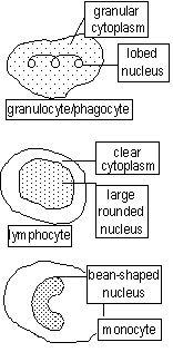
colourless, irregular (amoeboid) shaped, larger than RBC, nucleated, formed in lymph glands/ nodes, spleen and bone marrow, fewer than RBC (RBC:WBC=600-700:1)
Function-protect body by killing germs or foreign particles. Amount of WBC increase when fighting infections. WBC defend body in 3 ways:
1.phagocytosis-by granulocytes, bacteria engulfed by WBC > destroyed by lysosomes in WBC's cytoplasm
2.anti-toxin production-WBC contain anti-toxins which destroy toxins produced by bacteria (neutralize toxin's effects)
3.antibody production-lymphocytes produce antibodies (a protein) which gives immunity (ability of body to fight infections)[antibodies attack antigens (chemical substance of surface of cells) or attach to bacteria to immobilise it]
Leukemia-blood cancer, large amount of WBC in blood
Platelets
small round, oval or rod shaped bodies, enucleated, colourless, probably formed from red bone marrow
Function-Involved in blood clotting, release enzyme, thrombokinase
Blood Fluid-Plasma
structure-pale yellowish liquid, about 90% H2O in which contains a dissolved mixture of various substances.
Transports:-plasma proteins(serum albumin, globulin, fibrinogen, prothrombin)
-dissolved mineral salts (chlorides, bicarbonates, sulphates, phosphates, calcium, etc.)
-food substances (glucose, amino acids, fats and vitamins)
-excretory products (urea, uric acid, creatinine, etc.)
-CO2 (in HCO3- ,hydrogen carbonate ions)
-blood cells, platelets, antibodies, hormones, ions, heat, substances needed for blooding clotting
Clotting of blood (vit. K + calcium)
needed to stop 1too much blood being lost, 2pathogens from entering body via wounds. 1st step to healing of wound
Mechanism of clotting; Blood (of damaged blood vessel)>(exposure to air)>thrombokinase (produced by platelets + damaged tissues) Thrombokinase >(in presence of Ca salts)> prothrombin. Fibrinogen >(prothrombin)> insoluble fibrin threads>form mesh/clot, trapping blood cells>mesh shrinks>pale yellow serum (plasma w/o fibrinogen) squeezed out
Clot in blood vessels > heart attacks. Anti-coagulants prevent clotting in blood vessels(heparin, produced in liver)
Haemophilia-genetical disorder, affects blood clotting mechanism, substance needed for clotting absent, no clotting
Haemorrhage-loss of blood
A person can lose 1 or 2 liters of blood w/o ill effect, but if more is lost, in danger because
-low blood pressure, which slows down flow of blood round the body
-little amount of RBC, less oxygen transported
Blood vessels
Blood vessels; branched tubes which carry blood from heart to all parts of body, and returning blood to heart
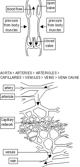
-Arteries -Capillaries -Veins
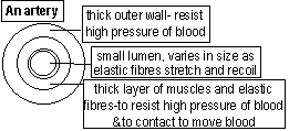
Arteries
-carry blood away from heart
-carry bright red oxygenated blood (except pulmonary + umbilical artery)
-largest-aorta, leaves left ventricle branches into arteries
-generally deep seated
start as large tubes, branching into smaller tubes, arterioles, which divide into capillaries
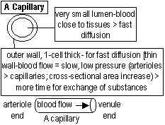
Capillaries
-small short tubes forming a network-large surface are for exchange of substances
unite to form a venules which unite to form veins before leaving organ
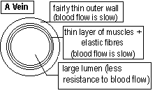
Veins
-carry blood to heart
-carry dark red deoxygenated blood (except pulmonary + umbilical vein)
-largest-vena cavae
-generally superficial (close to skin surface), esp. in limbs
-blood flow=slow (low pressure), valves prevent back flow of blood,
[movement of blood along veins assisted by action of skeletal muscle on veins (pressure on veins)
start as narrow tubes, joining up to form larger tubes
The Heart
-Considered as the centre of the circulatory system, collects blood and pumps it throughout body through blood vessels
-Features of pumping organ;1.made of cardiac muscle (don't get fatigue)
2.valves make blood flow in correct direction (atrium to ventricle)
3.septum prevents blood from mixing
-lies in chest cavity (to the left) between lungs, protected by chest wall muscles, ribs, backbone, sternum + diaphragm
-enclosed in pericardium-a membrane sac (protects heart)
-pericardial fluid in pericardium reduces friction when heart beats
-hollow, muscular organ, ~ 255-280g
-divided by septum into left and right side
-atria-smaller & have thinner walls (only collects blood and passes it on to ventricles)
-ventricles-bigger & have thicker walls (pump blood at high pressure)
-LV-thickest wall (3 times thicker than RV wall)-pumps blood to whole body at high pressure
(coronary arteries, arising from base of aorta, supply blood to heart muscles)
-valves prevent back-flow of blood (makes blood flow in correct direction, A's > V's) (bicuspid-2 flaps, tricuspid-3 flaps)
-cordae tendineae=cord-like tendons; attach valve flaps to muscular projections in ventricle walls
(prevent flaps from being turned back into atrium)
-flaps point downwards; permit easy flow of blood from atrium > ventricle
Heart Beat-Cardiac Cycle
-Pumping action due to systole (contraction) period + diastole (relaxation) period (atrial/ventricular systole/diastole)
-a heart beat takes about 0.8s. Adult; 70-76 heart beats per minute
-vigorous exercise + emotional condition increase heart beat
The Pulse
Each heart beat sets up a ripple of pressure, which passes along arteries & can be felt as a pulse as the arteries' muscular walls expand and contract. A pulse is produced after ventricular systole. Number of heart beat & pulse rate are similar. Pulse rate: same for all arteries (neck and wrist pulse) Pulse felt w/ finger-tips presses lightly over artery. (thumb not used as it has it's own pulse rate) Pulse rate varies from person to person, after exertion-high, rest-low
Blood Pressure
Force of blood exerted on walls of blood vessels. Blood pressure; arteries: highest-ventricular systole, diastole> decreases. Varies in different parts of body and from person to person. Average person-120-140mm/Hg systolic pressure and 75-90mm/Hg (80) diastolic pressure. Blood pressure measured by a sphygmomanometer. High blood pressure-hypertension (can occur after heavy exercise or when angry) causes heart to work harder > heart failure, can cause damage to kidneys and eyes and increase in risk of arteries tearing open. Stroke: artery that supply blood to brain ruptures
Double Circulation
Mammals-only vertebrae w/ 2 circulations-double circulation, blood passes through the heart twice.
Pulmonary circulation (low pressure circulation)-deoxygenated blood: RA > tricuspid valve > RV. RV contracts, blood > pulmonary artery > lungs. In lungs (alveoli) gaseous exchange occurs CO2 removed, O2 taken in. Blood oxygenated > pulmonary vein > LA
Systemic circulation (high pressure circulation)-main circulation. Oxygenated blood: LA > bicuspid valve > LV, LV contracts, blood > aorta which branches into arteries; supply pure oxygenated blood to organs. [renal-kidneys, hepatic-liver, coronary-heart, iliac-limbs] Veins collect deoxygenated blood>superior/anterior+inferior/posterior vena cavae>RA
Coronary Thrombosis
Heart depends on 2 coronary arteries to supply O2 to heart muscles (uses O2, 2nd most) Blood flow obstructed > heart muscles stop functioning > heart attack may occur.
Factors of obstruction;
i)coronary thrombosis-blood clot in arteries
ii)atherosclerosis/arteriosclorsis-deposition on yellow fatty patches under walls (atheroma) which narrow lumen
iiii)spasm-repeated contraction of muscles in coronary artery wall
A heart attack is often caused by a combination of the factors. If main coronary artery blocked, entire heart muscle may be deprived of O2 and nutrients > cells die. In early stages of heart attack, deposits of atheroma cause blood flow to become unusual > angina-a kind of pain in chest during strenuous exercise (warning of risk of a heart attack) Platelets begin to group over the atheroma > complete obstruction of coronary arteries. If unattended, an angina victim could die
Risk factors
Smoking-major factor of clotting + atherosclerosis. CO + other chemicals in cigarette smoke > damage lining of arteries
> atheroma form
High level of fat-(esp. cholesterol in food) major cause of atherosclerosis. Saturated fat is dangerous
High blood pressure-(hypertension + diabetes) contributes to atherosclerosis
Stress-emotional stress often raises blood pressure
Lack of regular exercise-exercise improves blood flow in heart. Lack of it causes blood to be sluggish >atheroma forms
Tissue Fluid
-colourless, watery fluid found in between cells, it is blood that leaks out of capillaries - (RBC+ plasma proteins + some WBC) (too large)
Function-bathes cells, keeps them in right condition & supplies substances needed by cells from bloodstream
Lymphatic system
too much tissue fluid > returns to capillaries or drained into lymphatic system, consisting of narrow lymph vessels containing a fluid-lymph. Lymph vessels eventually lead to veins > lymph enters blood stream. At intervals in lymph vessels > swellings = lymph nodes/glands. Lymph nodes-full of tiny spaces to filter lymph & produce lymphocytes to fight diseases (attack + destroy germs) thus acting as filters to pathogens. Main lymph glands-neck, armpit, groin
Respiration
Excretion
Homeostasis
The Eye
Nervous System
Chemical Control of Plant Growth
The Use and Abuse of Drugs
Diversity of Organisms
Nutrient cycles and Ecology
Parasitism
The Human Impact on the Environment
Reproduction in plants
Sexual reproduction in animals
Genetics
Cell Structure and Organisation
Enzymes
Nutrition
Transport in Plants
Support, Movement and Locomotion
Back to 'O' level notes index
Back to notes index


